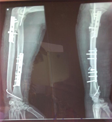

asymmetry of the distal radioulnar joint when compared to the other forearm 6.widening of the distal radioulnar joint on the frontal view 6.radial shortening may occur, and if greater than 10 mm, suggests complete disruption of the interosseous membrane.dislocation of the distal radioulnar joint.commonly at the junction of the middle and distal thirds.However, good quality orthogonal views are needed to identify and characterize displacement correctly.
DIFFERENCE BETWEEN MONTEGGIA AND GALEAZZI FRACTURE SERIES
Galeazzi fractures are classified according to the direction of radial displacement:Ī forearm series is usually sufficient for diagnosis and management planning.

Typically, Galeazzi fracture-dislocations occur due to a fall on an outstretched hand (FOOSH) and result in dorsal displacement of the radius (type I) if the axial load was applied to the forearm in supination or volar displacement of the radius (type II) if the forearm was in pronation 7. >10 degrees dorsal angulation >5 mm shortening significant comminution) 1.Galeazzi fractures are primarily encountered in children, with a peak incidence at age 9-12 years 3. In adults, it is estimated to account for ~7% of forearm fractures 3. Open reduction and internal fixation (ORIF) is considered when the fracture is unstable, and/or unsatisfactory closed reduction is achieved (i.e. The cast extends from below the elbow to the metacarpal heads and holds the wrist somewhat flexed and in ulnar deviation 4 - for those of you familiar with Australian rules football this position is reminiscent of the position adopted when holding a ball in preparation for a kick. The vast majority of Colles fractures can be treated with closed reduction and cast immobilization. Location of the medial fracture line: does it involve the radioulnar joint In addition to noting the presence of a fracture a number of features should be sought and commented upon: An associated ulnar styloid fracture is present in up to 50% of cases.Ī pronator quadratus sign is generally seen. If dorsal angulation is severe enough, a dinner fork deformity may be described.There is also usually impaction with resultant shortening of the radius. Dorsal angulation of the distal fracture fragment is present to a variable degree (as opposed to volar angulation of a Smith fracture). The fracture appears extra-articular and usually proximal to the radioulnar joint. The plain radiographic series often comprises an AP and a lateral view however, it is not uncommon for an oblique view to be included. Plain films usually suffice, although if there is a concern of intra-articular extension, then CT may be beneficial.

As such, in clinical practice, the use of the term Colles fracture with an appropriate description of any associated injuries is sufficient in most instances. One of the more popular is the Frykman classification system, although it fails to distinguish between Smith and Colles fractures as it is based on AP radiographs 2,3. Radiographic featuresĪ number of classification systems exist for distal forearm fractures. Most fractures are therefore dorsally angulated and impacted. The proximal row of the carpus (particularly the lunate and scaphoid) transfers energy to the distal radius, both in the dorsal direction and along the long axis of the radius. Most Colles fractures are secondary to a fall on an outstretched hand (FOOSH) with a pronated forearm in wrist extension (the position one adopts when trying to break a forward fall).


 0 kommentar(er)
